Speaker
Description
The SIRMIO (Small Animal Proton Irradiator for Research in Molecular Image-guided Radiation-Oncology) project [1] aims at developing a portable preclinical proton irradiator that can be installed at existing proton therapy centres. The clinical beam properties will be adapted to match requirements of small-animal irradiation using a dedicated energy degradation and focussing system [2]. Pre-treatment proton imaging will be performed for position verification and treatment planning. For compatibility of the imaging setup with synchrocyclotron-based facilities, where the high instantaneous particle flux exceeds detection capabilities of contemporary single particle tracking systems, we are developing a compact solution based on a miniaturized Timepix detector.
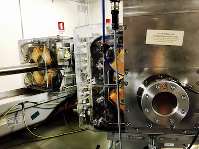
We tested the feasibility of our setup at the experimental beamline of the Trento Proton Therapy facility with a 70 MeV proton beam. A miniaturized radiation camera MiniPIX [3], based on the single particle tracking ASIC detector Timepix, was placed 1 cm behind a µCT calibration phantom consisting of a 10 mm thick solid water slab with 10 cylindrical tissue-equivalent inserts (16 mm length, 3.5 mm diameter). The reduced beam intensity (< 10,000 protons/second) assured the registration of individual protons for detailed event-by-event analysis despite the frame-based detector readout. The events were spatially binned and the median energy deposition within the 300 µm thick silicon sensor chip was calculated for each bin. It was converted to water-equivalent thickness (WET) of the traversed material using a FLUKA [4,5] Monte Carlo based and experimentally validated conversion curve. The obtained WET values for all inserts were divided by their geometrical length and compared to relative (to water) stopping power from literature [6].
For all insert materials, the WET was in good agreement with literature (differences < 3%) at an estimated imaging dose of 5 mGy. The spatial resolution was 0.3 mm. Increasing the phantom-detector distance from 1 to 5 cm resulted in more blurring and hence worse spatial resolution. Yet, even at an air gap of 5 cm, spatial resolution was better than 0.7 mm. The acquisition time per radiography was 15 – 20 min.
While spatial and WET resolution at an acceptable dose show the feasibility of our compact setup, the acquisition time is too long to image living samples. This shortcoming can be solved by replacing the detector by its successor model (Timepix3-based), which features event-based readout and can hence sustain much higher particle rates. Imaging time can then be reduced by about two orders of magnitude, allowing faster radiographic, and ultimately tomographic imaging.
Supported by ERC-Grant 725539.
[1] Parodi et al, Acta Oncol, 58 (2019)
[2] Gerlach et al, PMB, (2020, tentatively accepted)
[3] Granja et al, Nuc Inst Meth Phys Res A, 908 (2018)
[4] Böhlen et al, Nuclear Data Sheets 120 (2014)
[5] Ferrari et al, CERN-2005-10 (2005)
[6] Hudobivnik et al, Med Phys 43 (2016)

