Speaker
Description
In hadron therapy a highly conformed irradiation field is delivered to the target by moving the beam and modulating its energy. Treatment plans require precisely measured patients’ Stopping Power (SP) maps, which are presently extracted from X-rays tomographies, so introducing unavoidable uncertainties. A direct measurement of the SP maps using protons (proton Computed Tomography - pCT), could mitigate this source of errors potentially enhancing the precision of the hadron therapy.
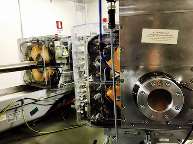
The Prima-RDH-IRPT collaboration built a 5x20 cm$^{2}$ field of view pCT system, suitable for pre-clinical studies, using a microstrip silicon tracker and a YAG:Ce calorimeter.
In this talk a detailed description of the apparatus, together with the measurement methodology, will be given. Tomographies of electron density calibration and anthropomorphous phantoms taken using the experimental beam at the Trento Proton Therapy center will be shown. Very good correlation between measured and expected relative SP has been obtained from the density phantom tomography with discrepancies less than 1%. Anatomical structures of the order of one millimeter are visible in the anthropomorphous head phantom image as well as details of a titanium spinal bone prosthesis and a tungsten dental filling. Furthermore, pCT tomographies of the head phantom taken with our device, when compared with x-CT images of the same object, evidence a significant reduction of artifacts induced by the prostheses.

